
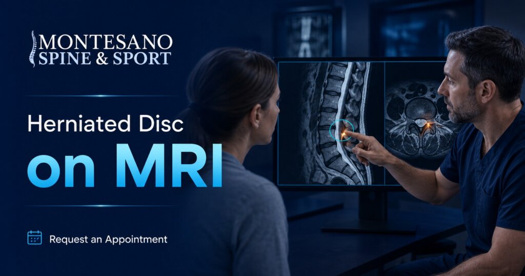
Quick Answer: A herniated disc on MRI appears as focal disc material extending beyond the normal disc space. Radiologists describe its level, direction, shape, and effect on nearby nerves. The finding must match the patient’s symptoms and examination before it guides treatment.
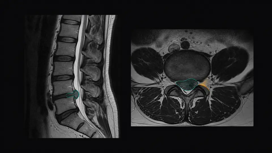
A herniated disc occurs when disc material moves beyond its normal boundary. MRI provides detailed views of discs, nerve roots, the spinal canal, and the spinal cord without ionizing radiation. This guide explains MRI appearance, report terminology, imaging indications, and how scan findings affect care.
Herniated Disc MRI
| Feature | What it means |
|---|---|
| Best imaging view | MRI shows discs, nerves, spinal cord, and surrounding soft tissues in multiple planes. |
| Typical exam | For suspected routine lumbar disc herniation, MRI is commonly performed without contrast. |
| Key report details | Level, side, location, morphology, nerve contact or compression, and canal or foraminal narrowing. |
| Common symptomatic levels | Lumbar herniations frequently involve L4-L5 or L5-S1, although any spinal level can be affected. |
| Main limitation | Disc abnormalities can be incidental and may not explain the patient’s pain. |
What Does a Herniated Disc Look Like on MRI?
On MRI, a herniated disc appears as a localized extension of tissue beyond the adjacent vertebral edges. On T2-weighted images, healthy hydrated discs are usually brighter than dehydrated discs, while displaced material may indent the thecal sac or narrow space around a nerve. Sagittal images establish the level and vertical extent. Axial images show whether the herniation is central, paracentral, foraminal, or far lateral and identify the affected side.
How Bulge, Protrusion, Extrusion, and Sequestration Differ
| Term | MRI description | Clinical meaning |
|---|---|---|
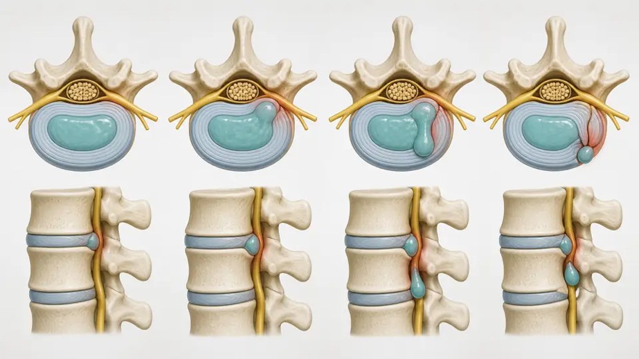
| Disc bulge | Broad extension of disc tissue, generally involving more than 25% of the circumference. | Often age-related and may be incidental. It can still narrow the canal or foramina. |
| Protrusion | Focal herniation whose base is wider than the outward portion. | May irritate or compress a nearby nerve, depending on location. |
| Extrusion | The outward portion is wider than its base, or disc material extends above or below the disc level. | More displaced material is present, but size alone does not determine symptoms. |
| Sequestration | An extruded fragment has lost continuity with the parent disc. | A free fragment may migrate and compress a nerve, yet some fragments shrink over time. |
An annular fissure is a split in the outer annulus fibrosus and can exist with or without a herniation. Disc desiccation, loss of disc height, endplate changes, and osteophytes describe degeneration rather than the herniation itself. Standardized terminology helps clinicians communicate, but wording may vary among radiologists and institutions.
Key Takeaway: MRI morphology describes the shape of displaced disc material, while location and nerve compression determine its likely clinical importance.
How to Read a Herniated Disc MRI Report
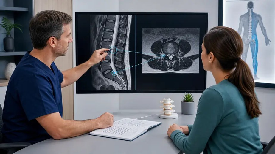
Read the impression first, then confirm details at each level. L4-L5, for example, identifies the disc. Central means midline, paracentral means just off midline, foraminal means within the nerve exit opening, and far lateral means beyond it. A report may say a disc contacts, displaces, impinges, or compresses a nerve root, although grading is not perfectly standardized.
Note central canal stenosis, lateral recess narrowing, and foraminal stenosis. These affect the main canal, the path of a traversing root, and the exiting root, respectively. Compare the report with pain distribution, strength, sensation, reflexes, and gait. A right L5-S1 herniation affecting S1 is more convincing when right-sided symptoms follow an S1 pattern.
Key Takeaway: The strongest MRI explanation is a level-and-side match between the compressed nerve and the patient’s neurologic findings.
Can an MRI Finding Be Present Without Symptoms?
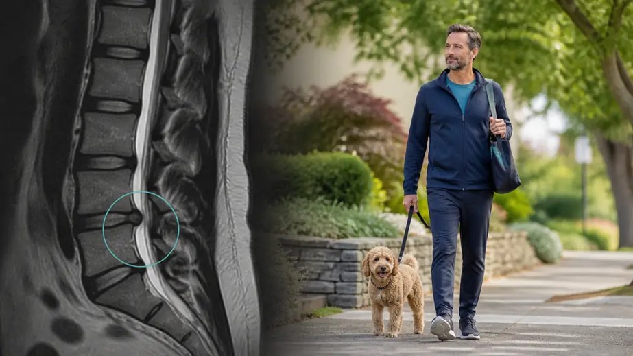
Yes. Disc findings are common without back pain. A systematic review found protrusions in about 29% of asymptomatic 20-year-olds and 43% of asymptomatic 80-year-olds. Disc bulges rose from 30% to 84% across those ages. MRI is therefore not a pain map or an automatic reason for surgery.
A small herniation can cause substantial symptoms when it irritates the right nerve, while a larger one may cause little pain if sensitive structures are spared. Chemical inflammation may also contribute to radicular pain. Clinical correlation helps avoid treating an incidental image.
Key Takeaway: An abnormal scan is clinically meaningful only when its anatomy plausibly accounts for the patient’s symptoms and examination.
When Is MRI Needed for a Suspected Herniated Disc?
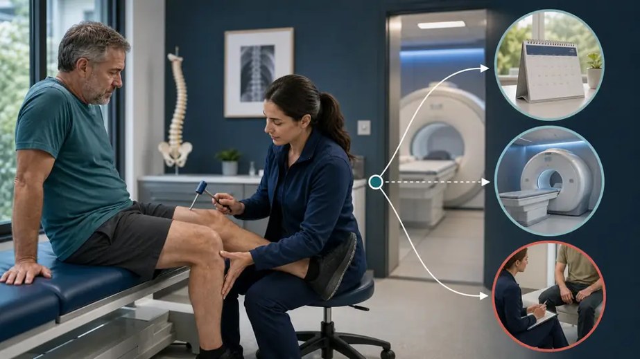
MRI is not routinely needed at the start of uncomplicated acute low back pain or sciatica. The American College of Radiology states that initial imaging is usually not appropriate when there are no red flags and no prior management. MRI becomes more useful when symptoms persist despite about six weeks of appropriate conservative care, when results would change an injection or surgical plan, or when serious disease is suspected.
Urgent assessment is needed for new bladder or bowel dysfunction, saddle numbness, rapidly progressive weakness, major trauma, or suspected cauda equina syndrome. Fever, immune suppression, cancer history, unexplained weight loss, or infection risk also changes the decision. Routine disc MRI usually needs no intravenous contrast. Contrast may help assess infection, tumor, inflammation, or selected postoperative questions. CT or CT myelography is an alternative when MRI is unsafe or limited.
Key Takeaway: MRI is most valuable when red flags are present, symptoms persist, or the result will directly change management.
What Happens After a Herniated Disc Is Found?

Treatment is based on symptoms and neurologic function, not the image alone. Most patients begin with activity modification without prolonged bed rest, appropriate pain medication when safe, and physical therapy focused on mobility and gradual return to function. An image-guided epidural steroid injection may help selected patients with persistent radicular pain, but it does not repair the disc.
Surgical consultation is considered for progressive weakness, cauda equina syndrome, persistent disabling pain, or a matching compressive lesion. Discectomy removes the fragment pressing on a nerve. Fusion or artificial disc replacement is not the default for every herniation and requires separate indications. MRI localizes the target, while benefits and risks depend on the complete clinical picture.
Key Takeaway: Most herniated discs are managed nonsurgically, while urgent or persistent neurologic compromise may justify surgical evaluation.
Best Practices for Discussing Your MRI
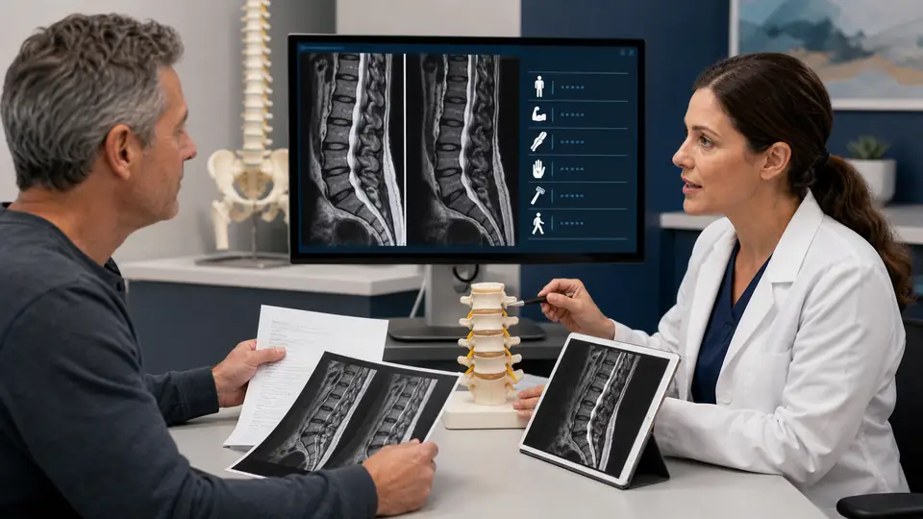
- Ask which specific finding matches your symptoms, side, and neurologic examination.
- Bring the images as well as the written report, especially when seeking a second opinion.
- Ask whether the finding is likely incidental, age-related, or responsible for nerve symptoms.
- Discuss what treatment would be recommended if the MRI were unavailable, then ask how the scan changes that plan.
- Seek urgent care for new bladder or bowel problems, saddle numbness, or rapidly worsening weakness.
Key Takeaway: A productive MRI discussion connects one specific imaging finding to symptoms, function, and a clearly stated treatment decision.
Common Mistakes When Interpreting a Herniated Disc MRI
Common mistakes include treating every bulge as dangerous, assuming a larger herniation must cause more pain, and ignoring the side or nerve level. Disc degeneration is not the same as an active herniation, and MRI usually cannot date an injury precisely. Repeat imaging is often unnecessary when symptoms and function are improving.
MRI cannot prove that a finding causes pain. Do not make medication, injection, or surgery decisions from one phrase in a report. A qualified clinician should review the study, examination, history, and prior images.
Key Takeaway: The most harmful interpretation error is equating an MRI abnormality with a diagnosis that automatically requires intervention.
Conclusion
A herniated disc on MRI is a focal displacement of disc material beyond its normal boundary. The report should state its level, location, shape, and effect on nerves. Protrusion, extrusion, and sequestration do not dictate treatment by themselves. Symptoms, examination, function, and imaging together determine whether observation, rehabilitation, an injection, or surgery is appropriate.
Key Takeaway: The right question is not only what the MRI shows, but whether that finding explains the patient’s symptoms and changes care.
Request an Appointment
Ready to discuss your symptoms or treatment options? Request an appointment with SpineDocs | Montesano Spine & Sport for an individualized evaluation.
SpineDocs | Montesano Spine & Sport
Phone: (888) 661-1408
Website: spinedocs.org
References
- American College of Radiology. ACR Appropriateness Criteria: Low Back Pain.
- RadiologyInfo.org. Spine MRI.
- American Academy of Orthopaedic Surgeons. Herniated Disk in the Lower Back.
- Fardon DF, et al. Lumbar disc nomenclature: version 2.0.
- Brinjikji W, et al. Imaging features of spinal degeneration in asymptomatic populations.
- Huang Z, et al. Value of imaging examinations in diagnosing lumbar disc herniation.
- Mayo Clinic. Herniated disk: diagnosis and treatment.
- North American Spine Society. Clinical guideline for lumbar disc herniation with radiculopathy.