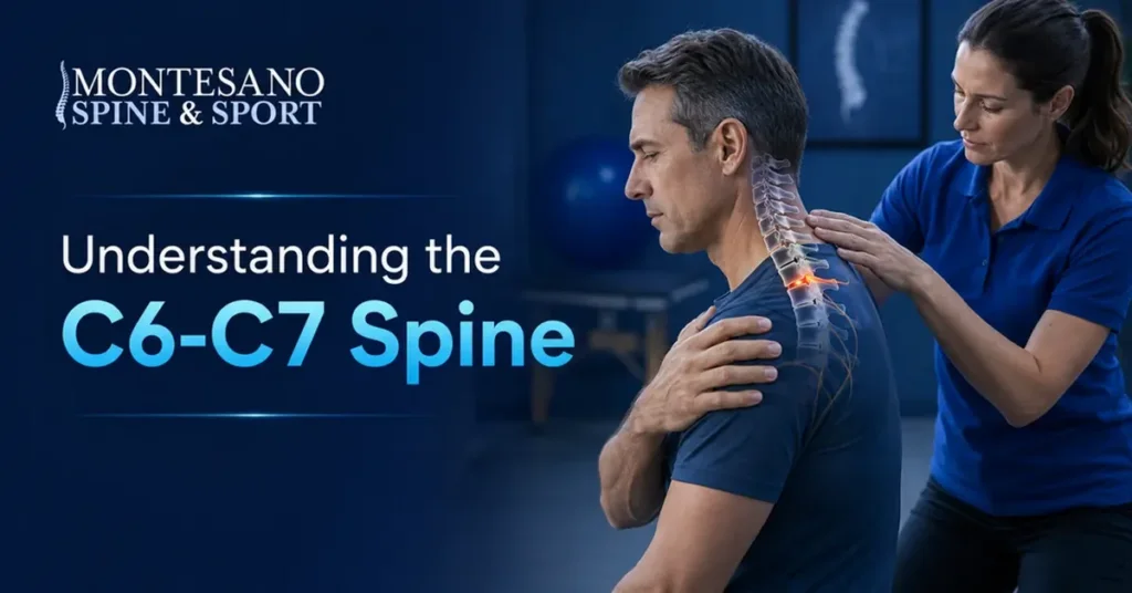
Quick Answer: The C6-C7 level is the lowest mobile disc space in the cervical spine and a common site of degeneration or herniation. Compression of the C7 nerve root may cause pain from the neck into the shoulder and arm, tingling toward the middle finger, and weakness in the triceps or pushing motion.
A finding at C6-C7 can mean anything from an incidental MRI change to a disc herniation causing arm symptoms or spinal cord compression. The right treatment depends on whether symptoms, examination findings, and imaging point to the same level, and whether there is weakness, loss of coordination, or persistent pain despite nonsurgical care.
C6-C7 Problems at a Glance
| C6-C7 issue | Common pattern | Typical next step |
|---|---|---|
| Disc degeneration | Neck pain or stiffness, sometimes no symptoms | Clinical exam and conservative care |
| C7 nerve-root compression | Arm pain, middle-finger tingling, triceps weakness | MRI plus neurologic examination |
| Spinal cord compression | Hand clumsiness, gait change, hyperreflexia | Prompt spine evaluation |
| Persistent symptoms after care | Pain or weakness limits daily function | Discuss injection or surgical options |
C6-C7 Anatomy and Function
C6 and C7 are the two lowest numbered cervical vertebrae. The disc between them cushions load and allows flexion, extension, and rotation. At this level, the C7 nerve root exits through the neural foramen, while the spinal cord passes through the central canal.
Age-related disc dehydration, bone spurs, a focal herniation, or narrowing of the foramen can irritate the nerve. Central narrowing can affect the cord. These changes may appear with degenerative disc disease or multilevel degenerative disc disease.
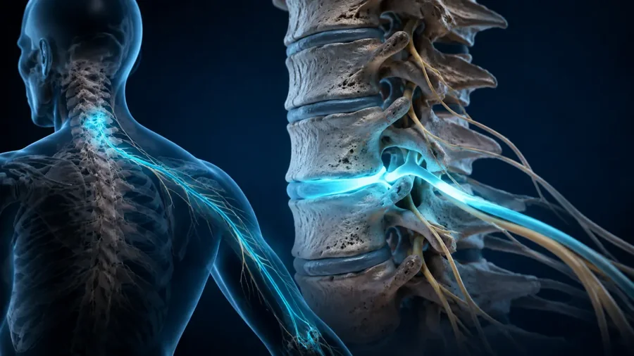
Key takeaway: C6-C7 problems can affect a nerve root, the spinal cord, or local neck structures, and those are clinically different problems.
C6-C7 Symptoms and the C7 Nerve Pattern
C7 radiculopathy may cause neck or shoulder-blade pain that travels down the back or side of the arm. Tingling or numbness can reach the middle finger. Weakness may involve the triceps, wrist or finger extension, grip, or pushing away from the body, and the triceps reflex may be reduced.
Not every person follows a textbook map. Shoulder, elbow, wrist, peripheral nerve, and paracentral disc protrusion problems can overlap. Cord compression may cause hand clumsiness, dropping objects, balance difficulty, leg stiffness, or changes in bowel or bladder control.
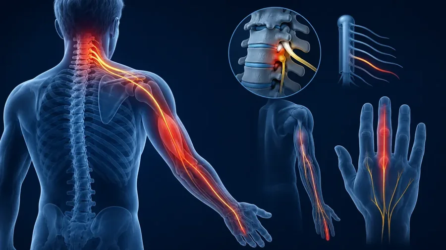
Key takeaway: The classic C7 pattern is useful, but diagnosis requires matching symptoms with strength, reflexes, sensation, and imaging.
How C6-C7 Problems Are Diagnosed
Evaluation includes symptom timing, aggravating positions, prior injury, gait, strength, reflexes, sensation, and tests that reproduce or relieve nerve tension. X-rays show alignment and motion. MRI evaluates discs, foramina, the spinal cord, and soft tissue. CT can better define bone, while electrodiagnostic testing may help when the level is unclear or a peripheral nerve disorder is possible.
A report that says “C6-C7 degeneration” is not a diagnosis by itself. The deciding question is whether the imaging finding explains the clinical pattern and whether neurologic function is stable.
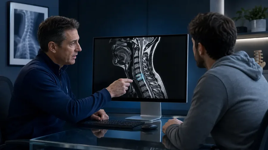
Key takeaway: The most useful diagnosis is a three-way match among the history, neurologic examination, and imaging.
Nonsurgical Treatment
Many patients improve with temporary activity modification, anti-inflammatory or other prescribed medication, and physical therapy focused on posture, mobility, nerve tolerance, and shoulder-blade and neck strength. Short-term traction may help selected patients. A cervical epidural injection can reduce radicular inflammation for some people but does not repair the disc.
Best practices include staying gently active, using a staged home program, tracking strength as well as pain, and reassessing if symptoms fail to improve. Avoid prolonged bed rest, forceful neck manipulation during active neurologic symptoms, and relying on an MRI image without a clinical examination.
Key takeaway: Conservative care aims to calm the nerve, restore movement, and preserve function while monitoring neurologic status.
C6-C7 Disc Replacement Versus Fusion
Surgery may be considered for progressive weakness, spinal cord compression, severe persistent radicular pain, or functional loss despite appropriate nonsurgical care. Anterior cervical discectomy and fusion removes the diseased disc and stabilizes the level. Artificial disc replacement also removes the disc but is designed to preserve motion.
Device-specific 10-year studies report high satisfaction and durable improvement after cervical disc replacement, with fewer secondary surgeries than fusion in some randomized comparisons. Those results do not apply equally to every implant, level, or patient. Severe facet arthritis, instability, osteoporosis, deformity, or anatomy unsuitable for the implant may favor fusion. Review total disc replacement or spinal fusion for a broader comparison.
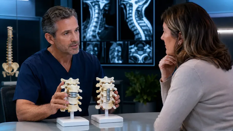
Key takeaway: Disc replacement and fusion both decompress nerves; candidacy, anatomy, goals, and surgeon experience determine which is more appropriate.
Risks, Recovery, and Expectations
Possible surgical risks include infection, bleeding, swallowing difficulty, hoarseness, nerve or spinal cord injury, persistent symptoms, device migration or wear, nonunion after fusion, and future adjacent-level disease. Recovery varies with the procedure, number of levels, health, work demands, and preoperative neurologic deficit.
Pain often improves before numbness or weakness, and some nerve symptoms can persist. Contact the surgical team promptly for worsening weakness, fever, drainage, breathing or swallowing difficulty, severe new pain, or loss of bladder or bowel control.
Key takeaway: A successful procedure improves the targeted problem, but nerve recovery and return to activity remain gradual and individual.
Conclusion
A C6-C7 MRI finding becomes meaningful when it matches a C7 nerve or spinal cord pattern. Most stable cases begin with focused nonsurgical care, while progressive neurologic loss or persistent disabling symptoms may justify surgery. A level-specific evaluation helps separate incidental wear from a treatable source of symptoms.
Request an Appointment
Ready to discuss your symptoms or treatment options? Request an appointment with SpineDocs | Montesano Spine & Sport for an individualized evaluation.